The Department of Biotechnology (DBT) supported National Cryo-EM Facility is located in the inStem building and is jointly managed by the Institute for Stem Cell Science & Regenerative Medicine (DBT-inStem) and the National Centre for Biological Sciences (NCBS-TIFR) on the Bangalore Life Science Campus. This is the first Cryo-EM that was installed in India for the analysis of biological samples. Currently the facility contains two electron microscopes including high-resolution transmission electron microscope (TEM) Tecnai T12 G2 spirit and high-resolution field emission scanning electron microscope (SEM) Merlin Compact VP as well as a sample, purification and preparation lab.
-
Transmission Electron Microscope or TEM (Tecnai G2 Spirit Bio-TWIN )
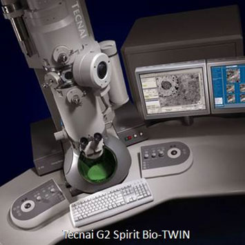
- High contrast, high resolution for 20 –120 kV operation Accelerating Voltage 20, 40, 60 80 100 120 kV
- Beam source: LaB6 filament.
- Vacuum system: Oil Diffusion (ODP) and Rotary for projection chamber; Ion (IGP) and Turbo vacuum pumps for column.
- +/-80˚ fully computer controlled Goniometer stage
- Optimized for 2D & 3D imaging of cells, cell organelles and soft matter
- Smart Tracking Position System for sample navigation Low dose mode available
- Cryo attachment for imaging of biological specimens and materials samples at sub-zero temperatures
- 4k x 2k GatanOrius side mount CCD camera.
- DM3 operation software (Gatan Digital Micrograph)
- 3D reconstruction software
- Holders: Standard single tilt; Gatan multiple specimen holder; Standard single tilt +/- 80˚ Tomography specimen holder; Gatancryo holder single tilt +/- 80˚ Tomography Cryotransfer Holder
-
Scanning Electron Microscope or SEM (The MERLIN Compact VP)
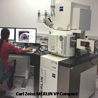
- Resolution: 0.8nm @ 15KV; 1.4nm @ 1 KV; 0.8nm @ 30KV (STEM mode)
- Probe current: 5 pA to 100nA
- Acceleration voltage: 0.02 V to 30 kV
- Magnification: 12 – 2,00,000 X
- Electron Emitter: Thermal (Schottky) field emission gun.
- Detection Modes
- Correlative Microscopy with Shuttle and Find.
- Cryo SEM and freeze fracture for imaging hydrated samples or for beam/vacuum sensitive specimens.
- EDAX for chemical composition analysis and element mapping.
- STEM: Low voltage optimized bright field, 4 quadrant dark field, and high angular dark field transmission imaging.
- Variable Pressure: High efficiency VPSE detector for pressure up to 60 Pa
- Secondary Ion: High efficiency in-lens SE detector Everhart Thornley Secondary Electron detector
- In-lens Duo: Switchable between on-axis in-lens secondary electron detection for highest surface sensitivity and energy selective backscattered detection for advanced materials contrasts.
- Specimen Stage: 5-Axes Motorized Eucentric Specimen Stage:X = 130mm; Y = 130mm; Z = 50mm; T = - 3º to 70º; R = 360º (continuous)
- Chamber: 330mm(Ø) x 270mm(h), 15 accessory ports for various options including CCD-Camera with IR-illumination
- Vacuum system: High vacuum mode and variable pressure mode
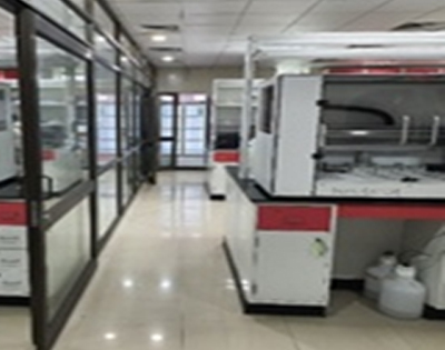
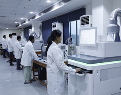
The facility began operations in 2018, and more than 66 users (both internal and external) have availed the electron microscopy facility.
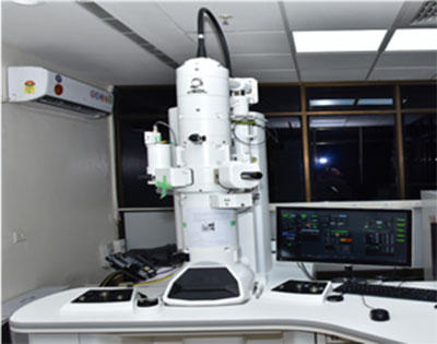 Transmission electron microscope (TEM) is used in a wide range of fields, such as biology and material sciences, to provide morphological and compositional information of samples. JEM1400 Flash is 120 kV TEM equipped with tungsten filament as an electron source and a highly-sensitive sCMOS camera. It can achieve high contrast imaging of biological specimens (including macro-molecular materials, tissue sections and viruses, etc.) and material science samples (polymeric and metallic nanoparticles, etc.) from low magnification (min. mag. 10X) to high magnification (max. mag. 1.2 MX) with resolution up to 0.38 nm.
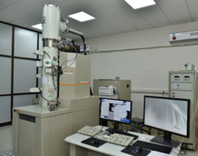 The JEM-2200FS is a field emission electron microscope that can be employed for biological and material science samples. It is equipped with a 200 kV field emission gun (FEG), a piezo-controlled goniometer, holders for cryo-observation and tomography, an in-column energy filter (Omega filter), and a Gatan camera (K2 summit direct detection camera). This instrument is utilized for high-resolution cryo-electron microscopy, zero-loss imaging, energy-filtered imaging, spectroscopy for elemental analysis, and tomography.
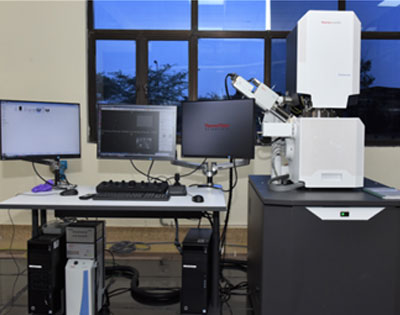 VolumeScope is a novel serial block-face imaging solution that enables excellent z-resolution from multi-energy deconvolution SEM combined with the efficiency of high-resolution cryo-electron microscopy in situ sectioning. The system offers the highest contrast and optimal SNR with in-lens and in-column detectors for HiVac, as well as dedicated detectors for optimal resolution in LoVac. The VolumeScope SEM unravels the complex 3D architecture of cells and tissues in their natural context, which is crucial for gaining an understanding of the structure-function correlation in biological systems.
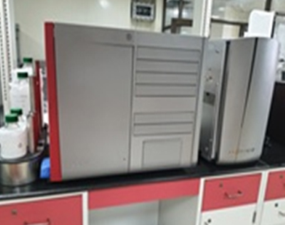
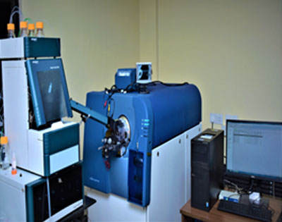 Mass Spectrometry enables the determination of the molecular masses of small molecules as well as of large biomolecules such as proteins and peptides. The SCIEX Triple TOF 5600+ mass spectrometer is a high-resolutionLC-MS/MSmass spectrometer with high sensitivity and high mass accuracy. The Triple TOF® 5600+ has both ESI and APPI ionization sources and is hyphenated with an ExionLC for LC-MS analysis of small molecules and also with an EksigentekspertnanoLC 425 for qualitative and quantitative proteomics analysis.
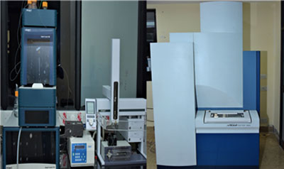 The facility also houses SCIEX TOF/TOFTM 5800 mass spectrometer with an EKSpot MALDI spotter for qualitative and quantitative proteomics analysis. The system is widely used for the identification of protein/ peptide, characterization of PTM, de novo sequencing, and relative quantification using iTRAQ, SILAC, TMT, etc.
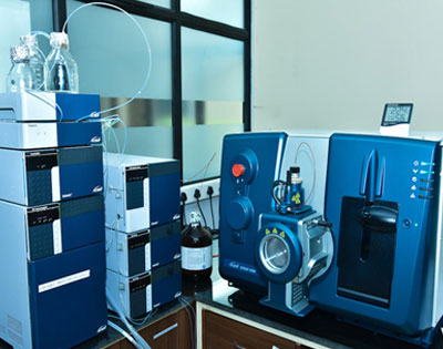 The facility is equipped with SCIEX QTRAP® LCMS/MS 6500+ system. It is the fastest and most sensitive QTRAP. It provides the best LOD's and LOQs to enable the detection and quantification of the widest scope of chemical compounds in the most challenging matrices. Improved polarity switching and MRM3 speeds allow faster chromatography and better throughput. The built in QTRAP functionality enables quantitative MRMs and qualitative QTRAP scans in the same injection to maximize throughput. It is best utilized for performing applications requiring ultimate sensitivity for ultra-low-level identification and quantitation of both small and large molecules and for confirmatory analysis of metabolites.
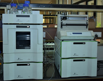 The facility provides services for cation exchange/high pH fractionation of complex proteomics samples using the Perkin Elmer FlexarTM HPLC system, which is coupled with an automated fraction collector for wide coverage of proteome.
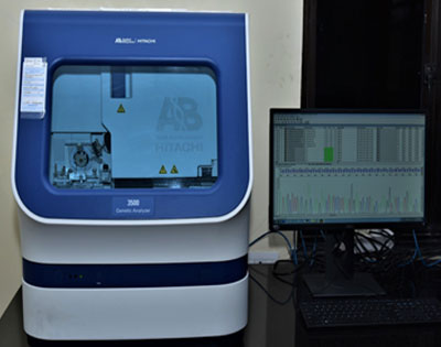 3500 Genetic Analyzer is a fluorescence based 8-capillary sequencing instrument which is based on Sanger DNA sequencing. The system is configured with 50 cm array and integrated software for instrument control, data collection, quality control, base-calling, and size-calling of samples. The system has a varied range of applications, like microbial sequencing, targeted DNA sequencing, epigenetic analysis, pathogen analysis, genetic disease research, SNP analysis, and biomarker analysis.
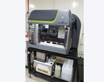
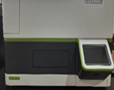
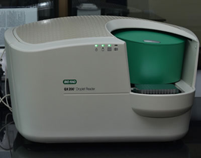 ddPCRTM is an advanced version of PCR that provides simultaneous clonal amplification and fluorescence-based quantification of nucleic acids. A typical reaction in ddPCRTM is partitioned into several thousands of water-in-oil droplets where individual reactions are carried out, providing absolute, precise, and reliable quantification. ddPCRTM has demonstrated usage in the detection of rare DNA targets, determination of copy number variations and measurement of gene expression levels.
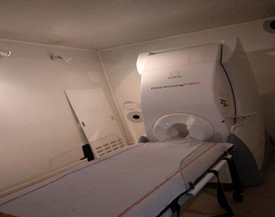 The objective of the facility is to perform MEG scans and analysis in persons with epilepsy (PWE) and issue the signed report for further procedures. At the MEG Resource facility, 2 PWEs are investigated every day. The scanning duration is 2-3 hours.
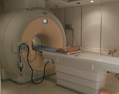
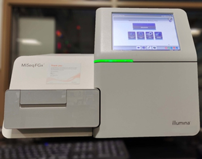 MiSeqFGx solution delivers a complete DNA-to-Data workflow for the analysis of forensic DNA samples. The solution begins with the ForenSeq™ DNA Signature Prep Kit, which includes all reagents required to prepare DNA libraries for sequencing. ForenSeq Universal Analysis Software delivers a powerful suite of forensic analysis capabilities including automatic detection of mixed DNA samples, generation of population statistics.
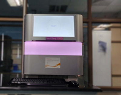
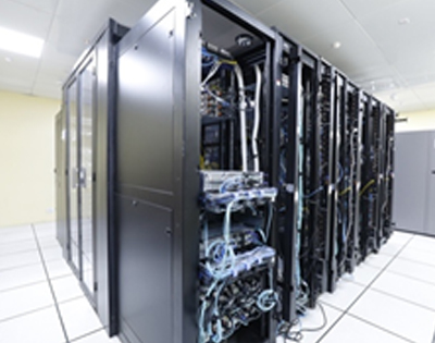 The facility also has Microarray setup for processing Illumina beads, a 300 TB Grid Scalar Central Data Storage platform (DDN), High performance parallel computing Clusters (HPPC Lab), computational labs for data analysis and also has a central Digital Library for accessing high impact journals through the DBT-Electronic LibraryConsortium (DeLCON) network. Flow cytometry, confocal imaging and tissue and bacterial culture are other basic facilities available